 This intensive training course is intended for academic or corporate staff wishing to improve their knowledge and skills regarding the acquisition and interpretation of images and data resulting from microscopy or cytometry techniques. As a four-and-a-half-day intensive event, it will combine theoretical classes (50% - limited to 10 trainees) and practical work (50% in sub-groups of 4 people maximum), interspersed with exercises designed to support trainees in their progress.
Employee, TRANSGENE SA
OBJECTIVES
- Acquire theoretical foundations in light and electron microscopy, cytometry and data analysis
- Understand the possible applications for each technology
- Understanding the practical aspect of these methods
PUBLIC & MODALITIES
- Public Researchers, engineers, and technicians wishing to familiarise themselves with fluorescence imaging techniques
- Staff numbers: Maximum of 10 trainees, practical sessions in sub-groups of maximum 4 trainees with 1 instructor per sub-group
- Evaluation: Throughout the training, practical exercises with solutions will allow the trainee to assess their acquisition of knowledge. Trainees' learning outcomes will be assessed at the end of the training and a training certificate will be issued.
- Trainees' equipment: It is advisable to bring your own laptop.
PROGRAMME
The course will emphasise the basic knowledge required to understand the different techniques and potential applications. Practical work will demonstrate the advantages and limitations of the various systems.
- Flow cytometry: principles, components of a cytometer, fluorescence and imaging flow cytometry
- Applications in flow cytometry: cell cycle, apoptosis, phenotyping, cell interaction, imaging flow cytometry, cytokine assay, metabolic probes, cell sorting
- Microscopy: the different types of light microscopy (bright-field, phase contrast, fluorescence?)
- Fluorescence microscopy: labelling, biosensors and applications
- The sample under the microscope: light/sample interaction and sample preparation
- The different types of fluorescence microscopes: performance, advantages and disadvantages
- Overview of sample preparation methods for transmission electron microscopy (TEM)
- Understand the basic principles of transmission electron microscopes
- Principles of scanning electron microscopes (SEM)
- SEM sample preparation
- Overview of data analysis
- Application to image analysis
Practical work (60 %) – Carried out in small groups on various equipment at the Toulouse imaging platform (widefield, confocal, multiphoton, SPIM microscopes, etc.)
- Advantages and limitations of the different systems
- Acquisition and analysis of images under the best conditions
Possibility of carrying out the practical work, for educational purposes, on samples brought in by trainees
EQUIPMENT & PERSONNEL
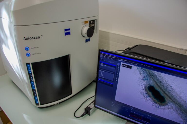
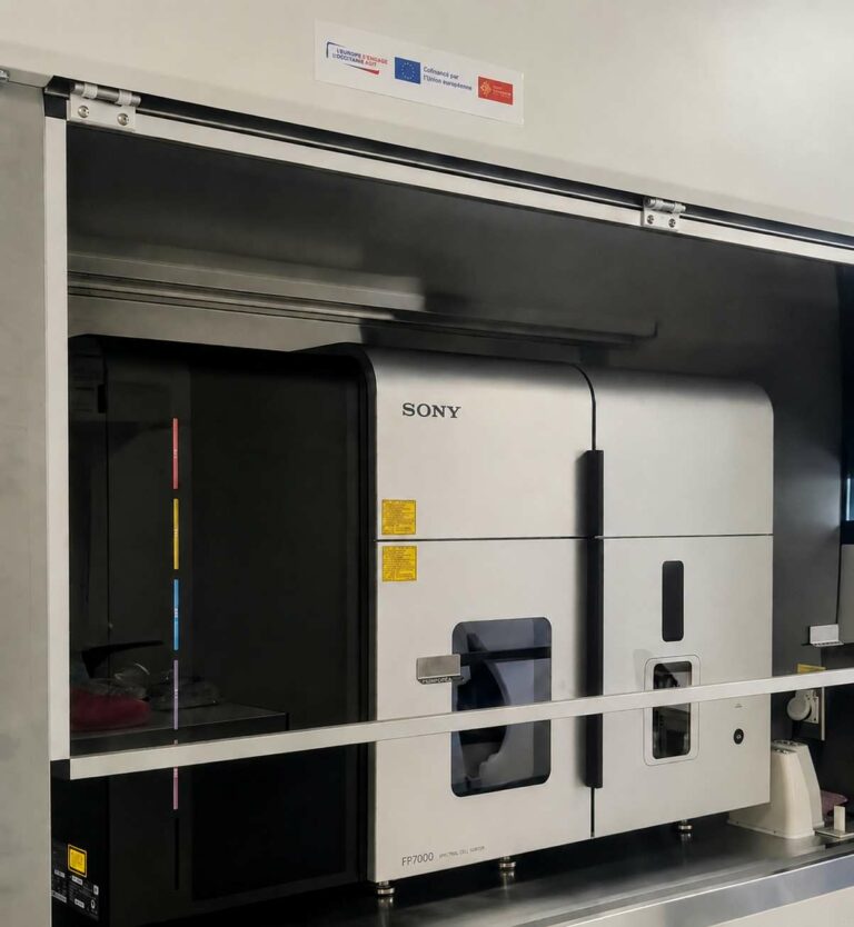
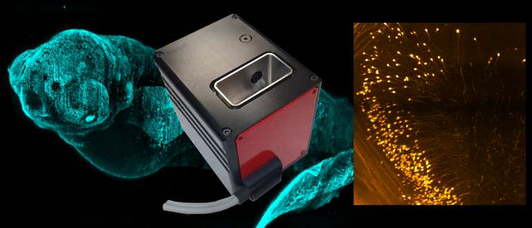
- Widefield, confocal, multiphoton, light-sheet microscopes, cytometers analysers, sorters, cytometers imaging, transmission electron microscopes, scanning electron microscopes. The equipment used is listed on the website (TRI).
- Engineers from the following research institutes: Agrobiosciences, Interactions and Biodiversity Research Federation; Biology and Biotechnology for Health Research Federation
Contact and registration
- For further information, contact: Jacques Rouquette
- Applications on the CNRS website
Qualiopi and AFAS ISO 9001 certified training