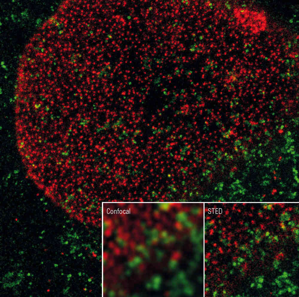


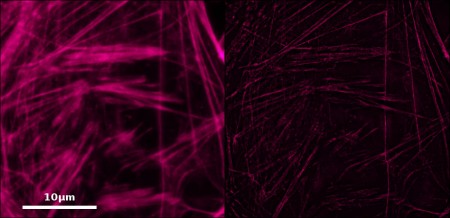
 In order to overcome the resolution limits imposed by laws of optics and thus to achieve higher resolutions, various fluorescence microscopy technologies have recently been developed. Grouped together under the terms «super-resolution» microscopy or «nanoscopy», they make it possible to localise, in vivo, right inside cells, compounds on the single-molecule scale (lateral resolutions of the order of 10 to 90 nm). These technologies, which rely either on principles similar to pointillism (PALM/STORM) or on structured illumination (SIM) or laser depletion (STED), earned their authors, E. Betzig, S. Hell and W. Moerner, the Nobel Prize in Chemistry in 2014.
Conducting competitive research across many fields of biology now requires access to super-resolution microscopy, which is why TRI is developing a super-resolution microscopy hub that benefits from these various approaches and is open to the entire scientific community.
Industrial Equipment
- dSTORM and STED type equipment at the CPTP. The former develops single-colour dSTORM approaches in TIRF microscopy, using a 400 mW 640 nm laser. As for the second, the STED, its originality lies in the fact that it is the only super-resolution microscope allowing work on 3D tissue sections, thereby providing access to human physiopathology with very high resolution (30 nm).
- Similar equipment, the N-Storm, is operational within the platforms united under the common LICT (CBD / CBI) banner. It uses the same technologies as the previous ones but allows for observations in a wider variety of wavelengths.
- A Zeiss ELYRA PS1 type equipment has been installed at the I2MC site. This super-resolution microscope uses a concept of nano-interaction analysis at the level of contacts between different cell types or between cells and the extracellular matrix. Thanks to this equipment, it is possible to analyse the same sample using the three technologies mentioned previously, SIM and STORM or PALM, in order to obtain an extremely detailed result, allowing single molecules to be localised with precision.
R&D technology
Two microscopes under development, based on a technique coupling 3D structured illumination (SIM) and localisation, are operational at the LITC site. Integrating the latest innovations in structured illumination imaging from internationally renowned microscopy laboratories (IPHT – Germany, Institut Fresnel – France, ISMO – France, Janelia Lab – USA), they make it possible to overcome the experimental constraints encountered in conventional super-resolution equipment. Technological support from the French companies Oxxius (laser technology) and AbbeLight (supercritical 3D localisation) will be in place over the course of 2016.
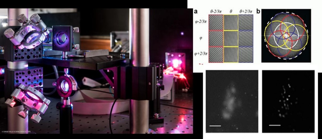
These items of equipment may be coupled with existing techniques such as optomanipulation:
- Conventional 2D TIRF-SIM and 3D SIM at a temporal frequency of 20 frames per second. This technique will thus enable super-resolution imaging of rapid phenomena such as mitosis and endocytosis,
- Fast 2D non-linear SIM enabling a resolution of 50nm using photo-activatable GFP molecules (acquisition rate: 5 frames / s). It will be dedicated to cytoskeleton and chromatin imaging (available in spring 2016),
- Single-objective multicolour isotropic 3D SIM imaging to democratise super-resolved isotropic imaging using a conventional microscope stand (available at the end of 2016),
- 2D SIM TIRF imaging combined with 3D localisation by supercritical angle fluorescence, in collaboration with AbbeLight (implemented in July 2016). It will achieve a resolution close to 20nm in Z,
- 3D SIM reconstruction via a new SIM deconvolving method adapted for live tissues (BLIND SIM), not available through commercial algorithms.
* « From microscopy to nanoscopy«, M. Moreau, P. Cochard, m/s no. 12, vol. 30, December 2014