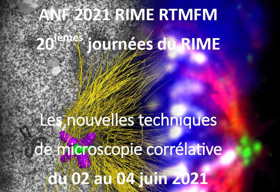

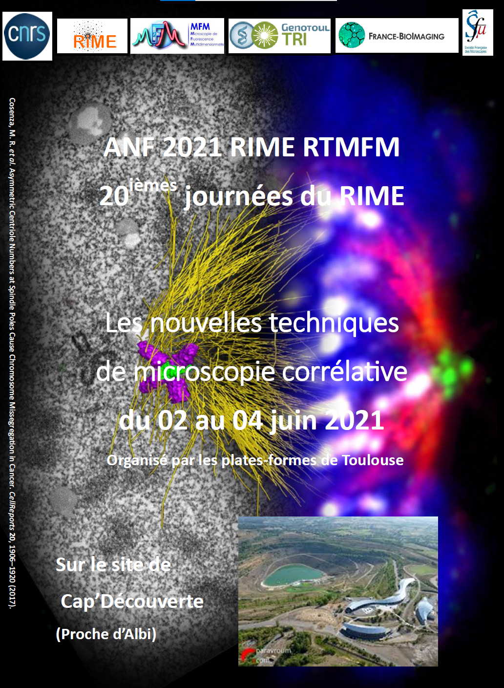
This 20eme edition of the National Training Action (ANF) in microscopy on «new correlative microscopy techniques» brings together technicians, engineers and researchers from both the public and private sectors wishing to deepen their knowledge of correlative microscopy. This two-day training course (3 days taking logistical aspects into account) will take place at the Cap’Découverte site (near Albi) to ensure compliance with health restrictions. Divided between plenary sessions, a poster session and workshops, this training will present the various correlative microscopy techniques, whether in terms of techniques or instruments. Traditional optical and fluorescence microscopy, scanning and transmission electron microscopy, NanoSIMS, X-ray fluorescence and synchrotron techniques will thus be covered. Enrolments before 15 April.
Functional photon microscopy imaging has taken a predominant place in biology, as it allows both the localisation and the measurement of the dynamics of molecules of interest in living cells, tissues or organisms, particularly in developmental biology. It is therefore a form of microscopy that makes it possible to work in vivo, but lacking the resolution to explore the entire cellular ultrastructure directly.
Electron microscopy, for its part, offers sub-nanometric resolution and a multidimensional scale, providing contextual information compared to optical microscopy. Nowadays, it is undergoing significant methodological development, notably through sample preparation and preservation techniques, and the development of electron cameras—particularly direct electron detection cameras—which bring greater and non-negligible resolution in cryo-EM, where samples are not chemically stained.
The need for precision in results regarding morphology or cellular immunocytology at the sub-nanometre scale is only increasing considerably in research. More than ever, most microscopists must integrate this aspect and find the appropriate solutions.
With the evolution of cryo-systems, 3D and image analysis software, new microscopy techniques are emerging. Added to this is the significant development of super-resolution techniques in light microscopy, which must be integrated into correlative microscopy approaches in order to bridge the resolution gap between light and electron microscopy. These developments pose new challenges both in terms of sample preparation and in terms of image alignment and registration.
During this training course, several correlative method approaches aimed at building multi-scale imaging of living systems will be covered, involving multiple technologies including electron microscopy and light microscopy, as well as AFM, X-ray fluorescence, cellular and tissue X-ray tomography, IR spectromicroscopy, and NanoSIMS.
Information and registration