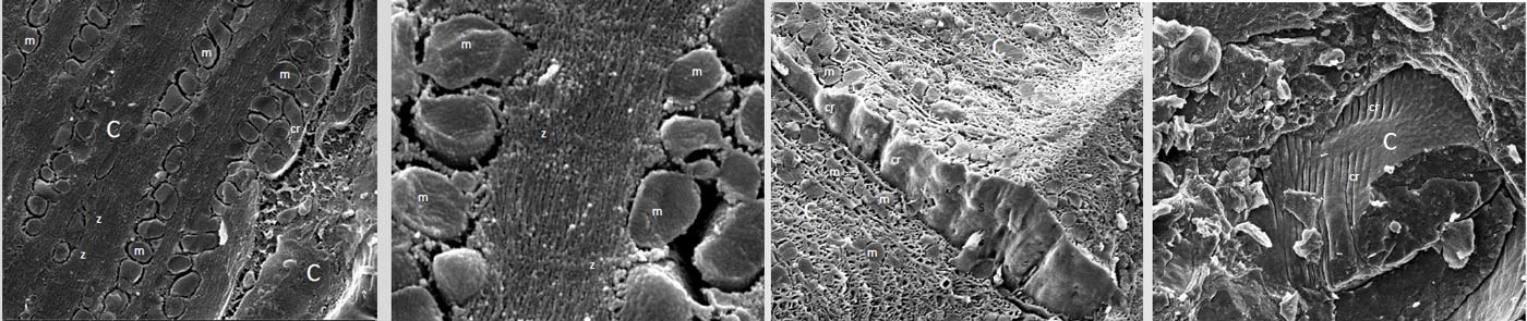

Unlike photon microscopy, electron microscopy requires a heavy pre-treatment of the samples to be observed. The goal for electronic platforms is therefore to propose methods of observing samples as close as possible to their native state. The CMEAB platform has recently developed a method of sample preparation by cryofixation that better respects the integrity of the sample.
The cryofixation of samples is a very delicate technique that must be performed under optimal conditions to avoid the formation of water crystals that significantly alter the ultrastructure of the samples. To do this, high-pressure cryofixation is currently the only technique that allows samples to be cryofixed without the use of protective coatings (cryoprotectants) whose thicknesses, approaching 200 μm, alter the appearance of the latter.
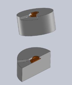
For this reason, the CMEAB platform chose high-pressure cryofixing as the preferred technique to prepare samples for observation after cryo-fracture in MEB. The technological hurdle encountered was the lack of an adapter allowing the fixation of the cupules of the high-pressure cryo-fixer. The objective within the RCCM working group was therefore to manufacture an adapter that would:
- the fixation of the cupules of the high-pressure cryostat (EM-ICE or HPM100, Leica),
- to be attached to the support of the cryo-preparation modules in MEB (Alto, Gatan or PP300 and PP3010T Quorum).
This technique has been tested and applied for the successful observation of the ultrastructure of mouse cardiomyocytes.