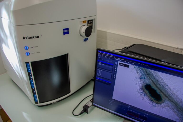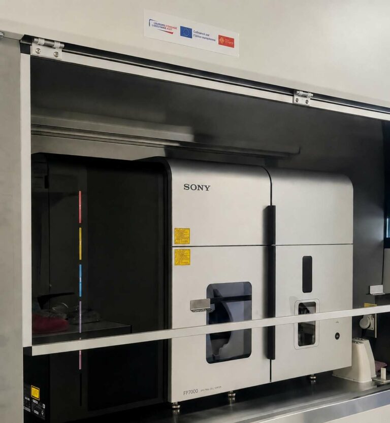


Living-sample analysis technologies in the field of human class 3 pathogen research have long been constrained by health and safety limitations. For the first time, engineers at the IPBS have brought together within a single biosafety level 3 laboratory unique imaging and flow cytometry tools that make it possible to work with live infected small animals, tissues and cells. This combination has been made possible through the adaptation and creation of state-of-the-art containment equipment, in collaboration with specialised equipment manufacturers. All of these technologies and expertise aim to drive ambitious and innovative research programmes in the field of infectious diseases.
- Check the dedicated page
- Here are a few photos taken on the platform
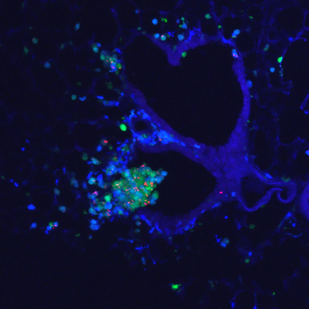
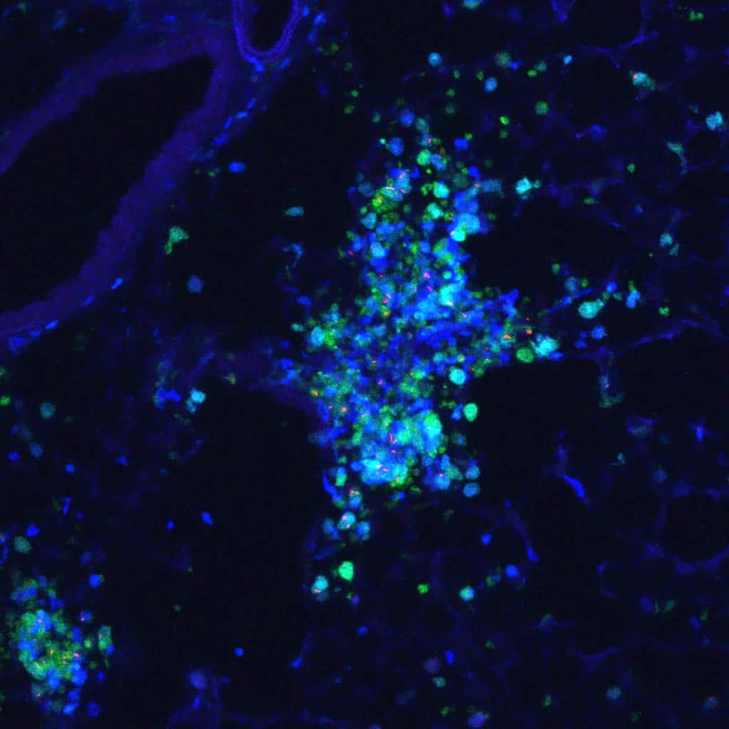
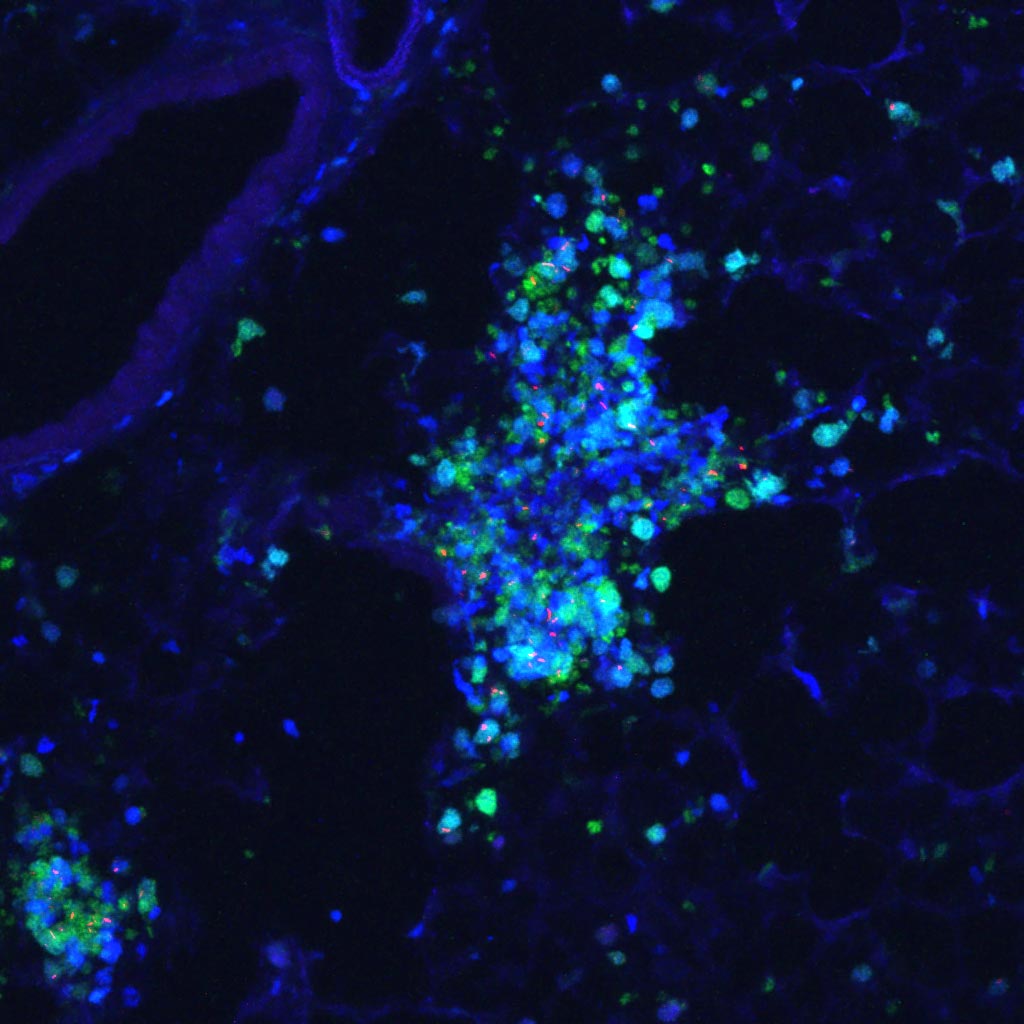 Lung of Macblue x LysM-GFP transgenic mice infected with Mycobacterium tuberculosis. Blue , macrophages and monocytes. Green neutrophils and monocytes. Red : M. tuberculosis. Credits: IPBS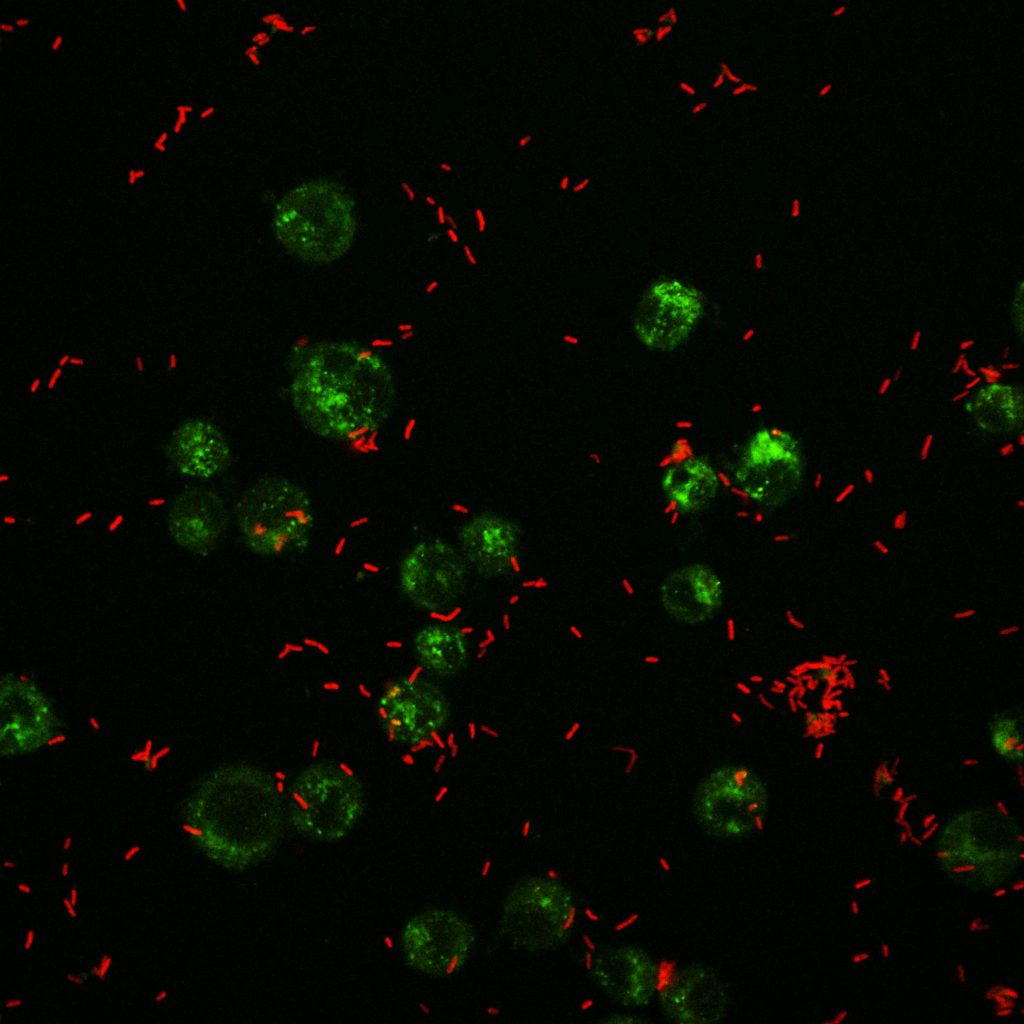
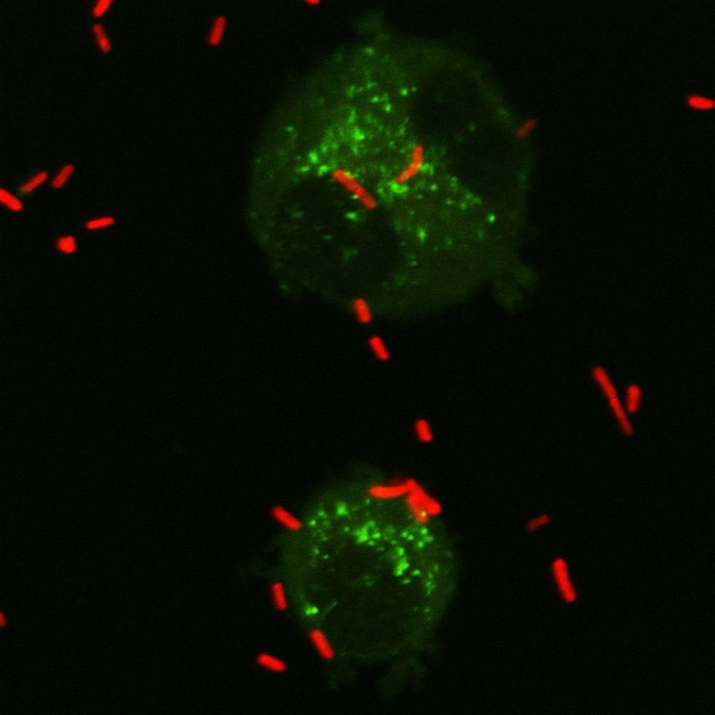 Alveolar macrophages infected by Pseudomonas aeruginosa (GreenFluozin labelling of intracellular zinc andRed) Pseudomonas aeruginosa – Credits: Danillo Pietretti, IPBS.