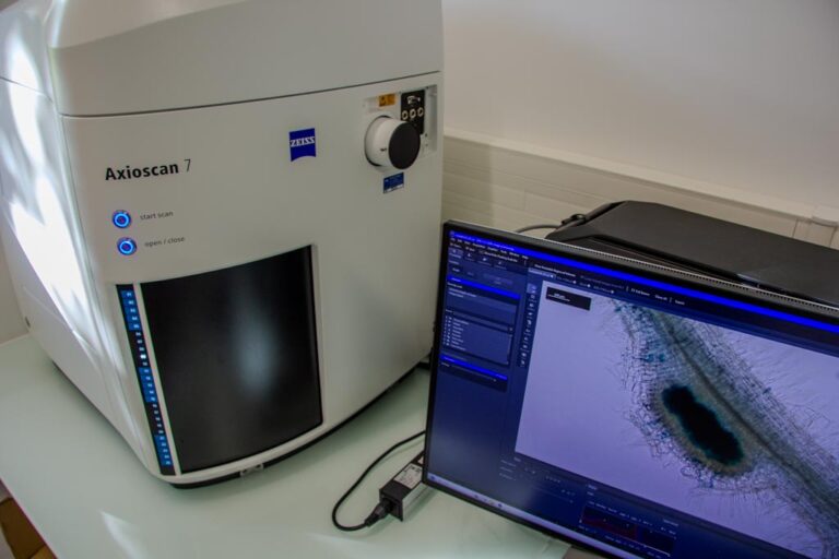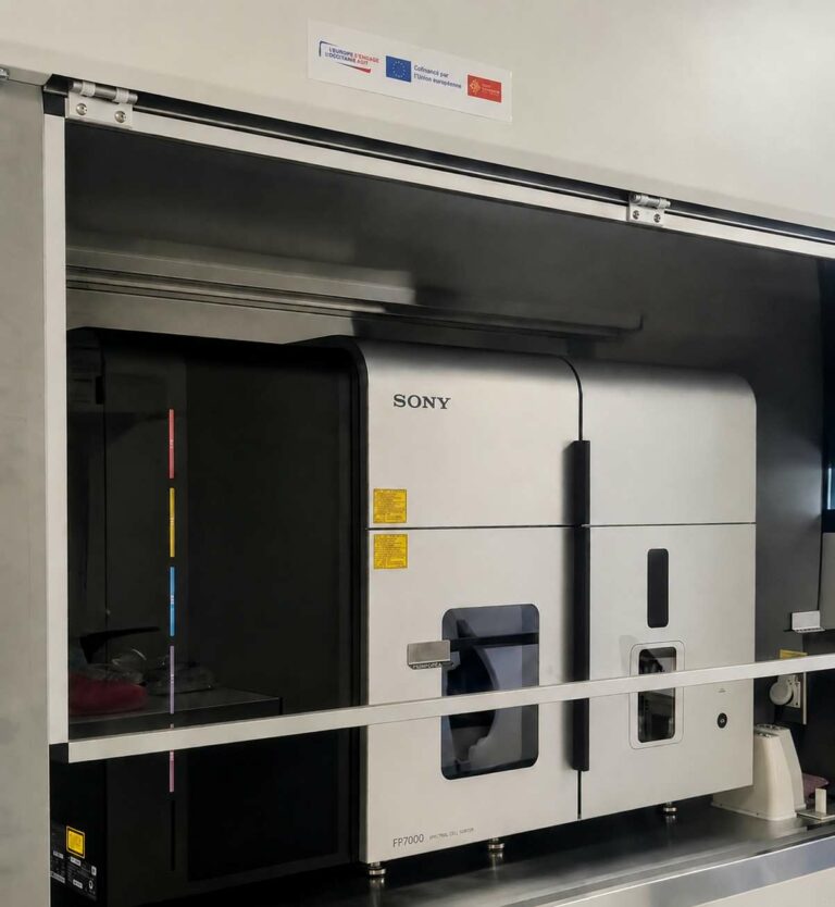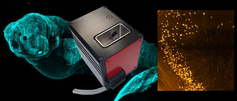


 The coupling of confocal imaging with live micromanipulation techniques enables a multi-scale approach to the study of l‘set of laws that determine the form and structure of tissues, the morphogenesis. The study of the forces locally exerted by a cell can then be integrated and analysed in relation to the dynamics of the cells making up the tissue as a whole, enabling a multi-scale biophysical model of this morphogenesis to be developed... It is the development of these new techniques that has made it possible today to discover that when a cell enters into apoptosis or cell death, it locally deforms the tissue into which it is integrated.
The LBCMP has implemented a laser nano-surgery technique (Ropper Scientific/Leica) on the TRI / CBI site, combined with 4D image analysis of Drosophila tissues. Nano-surgery is carried out at the subcellular scale using a pulsed laser beam. For example, it makes it possible to section cellular cytoskeleton filaments or cell junctions with extreme precision within a tissue without inducing significant heating, due to the very short duration of the laser pulse. Thanks to this technique, Magali Suzanne's team (CBI) has revealed the surprising behaviour of cells apoptotic (in the cell death phase) [1]. These cells exert temporary forces on their neighbours along the epithelium and generate new mechanical properties, inducing step-by-step the formation of a fold along an epithelial monolayer (fig. 1).
![A new active role for apoptotic cells in the formation of apoptosis-dependent epithelial folds (figure reproduced from [2]).](https://trigenotoul.com/wp-content/uploads/2015/04/figure-papier.jpg)
![Fig. 2: (a) Example of morphological analysis of fold cells in the control (ctl) context and upon apoptosis inhibition (Q-VD). (b) Measurement of relaxation time after laser nanosurgery in a control (DMSO) context and upon apoptosis inhibition (Q-VD). Figure modified from publication [1]](https://trigenotoul.com/wp-content/uploads/2015/04/figure2mortcellulaireolh.jpg)
In this process, image analysis and laser nano-surgery made it possible to quantify neighbouring cells mechanically and morphologically in the absence or presence of apoptotic tension. The relationship between the morphology of the cells forming the folds and their surface tensions measured by nano-surgery (Fig. 2) could thus be verified.
These data supported the idea that the force produced by the apoptotic cell can induce an increase in surface tension forces at the apex of neighbouring cells, thereby forming the future folds. As proved by a biophysical model and a genetic model in which myosin II was temporarily inhibited, both of these forces depend on this molecular motor.
Contact:
- Magalie Suzanne (Team Apoptosis-dependent morphogenesis, CBI)
- Thomas Mangeat (TRI / CBI Platform)
Resources used:
Publications
[1] B. Monier, M. Gettings, G. Gay, T. Mangeat, S. Schott, A. Guarner, M. Suzanne (2015). Apico-basal forces exerted by apoptotic cells drive epithelium folding. Nature, 518 (7538): 245-8.
See also: [2] Vasquez, C.G., Martin A.C. (2015) News and views, Cell biology: Death drags down the neighbourhood. Nature, 518 (7538):171-3.