

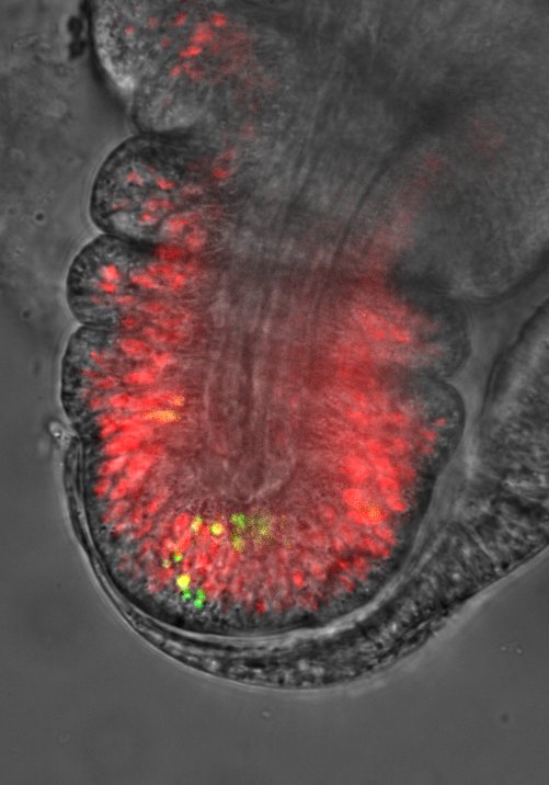
Apoptosis, or programmed cell death, has long been considered a simple mechanism for eliminating surplus cells. Today, we know that its role goes far beyond this, conditioning tissue patterning. Cells about to die thus transiently deform the tissue surrounding them and contribute to its final morphology. The team led by Magali Suzanne (CBI – UT3/CNRS), with the contribution of the LITC platform, explains this phenomenon, which involves the cell nucleus in the generation of mechanical force, in a study published in the journal Developmental Cell.
To understand the detail of the mechanism, it was necessary to observe the phenomenon in real time, at a very high frame rate in order to observe any movement. The spinning disc perfectly addressed this issue by enabling the capture of an image every second of an area focused on the cell nucleus. Two hypotheses had been considered by the researchers: either the nucleus is stabilised at the basal pole of the cell in order to resist the force generated by the contraction of the myosin cable to which it is attached, or it is not stabilised and its large volume is sufficient to provide the resistance necessary for force transmission. Spinning disk microscopy made it possible to decide in favour of the second hypothesis.
| Both films show the movement of the nuclei respectively: a zoom with nucleus tracking shows that the apoptotic nucleus (track1) is less mobile than the non-apoptotic nuclei (2 to 5) | |
- Details of the mechanism on the CNRS website
- Contact: Magali Suzanne
- Publication references : Mechanical Function of the Nucleus in Force Generation during Epithelial Morphogenesis – Ambrosini A, Rayer M, Monier B, Suzanne M – Developmental Cell Published online: June 13, 2019. DOI : https://doi.org/10.1016/j.devcel.2019.05.027