Combining alternating theoretical lectures (50 %) and practical sessions (50 %), this intensive training course is designed for company personnel wishing to improve their knowledge and skills regarding the acquisition and interpretation of images on various types of microscopes and cytometers. Taking place over 4 and a half days, this intensive event will combine lectures (limited to 10 trainees), practical sessions (in sub-groups of a maximum of 4 people with one or two instructors) and will be interspersed with exercises designed to support trainees in their progress.
OBJECTIVES
- Gain theoretical foundations in light and electron microscopy, cytometry and data analysis,
- Know the possible applications for each technology,
- Understanding the practical aspect of these methods
PUBLIC & MODALITIES
- Public Researchers, engineers and technicians wishing to discover all of these technologies
- Prerequisite: None, except for a clearly stated interest in in vivo optical imaging
- Staff numbers: Maximum of 10 trainees, practical sessions in sub-groups of maximum 4 trainees with 1 instructor per sub-group
- Evaluation: Throughout the training, practical exercises with solutions will allow the trainee to assess their acquisition of knowledge. Trainees' learning outcomes will be assessed at the end of the training and a training certificate will be issued.
PROGRAMME
The course will emphasise the basic knowledge required to understand the different techniques and potential applications. Practical work will demonstrate the advantages and limitations of the various systems.
- Flow cytometry: principles, components of a cytometer, fluorescence and imaging flow cytometry
- Applications in flow cytometry: cell cycle, apoptosis, phenotyping, cell interaction, imaging flow cytometry, cytokine assay, metabolic probes, cell sorting
- Microscopy: the different types of light microscopy (bright-field, phase contrast, fluorescence?)
- Fluorescence microscopy: labelling, biosensors and applications
- The sample under the microscope: light/sample interaction and sample preparation
- The different types of fluorescence microscopes: performance, advantages and disadvantages
- Overview of sample preparation methods for transmission electron microscopy (TEM)
- Understand the basic principles of transmission electron microscopes
- Principles of scanning electron microscopes (SEM)
- SEM sample preparation
- Overview of data analysis
- Application to image analysis
EQUIPMENT & PERSONNEL
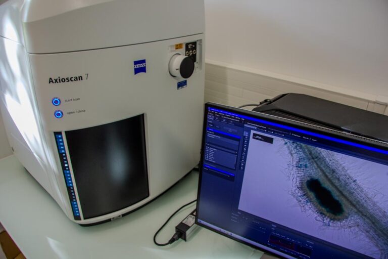
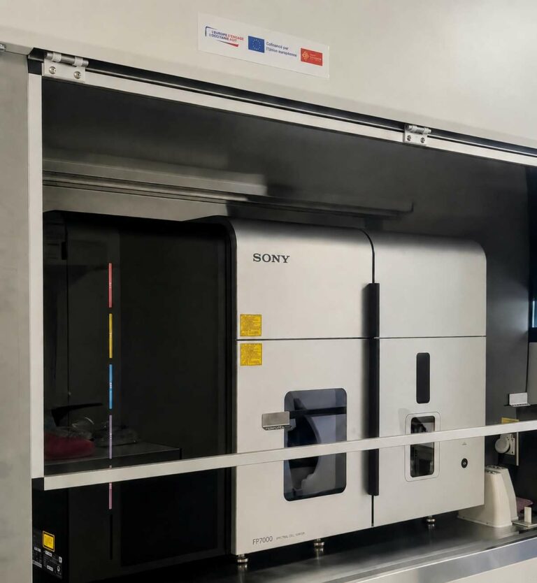
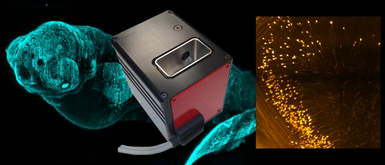
- Widefield, confocal, multiphoton and light-sheet microscopes, cytometers analysers, sorters, cytometers imaging, transmission electron microscopes and scanning electron microscopes,
- Engineers from the following research institutes: Agrobiosciences, Interactions and Biodiversity Research Federation; Biology and Biotechnology for Health Research Federation,
- The trainee is advised to bring their own laptop.
Contact and registration
- For further information, contact: Jacques Rouquette
- Applications on the CNRS website