 This intensive training course is aimed at academic or corporate staff wishing to improve their knowledge and skills regarding the acquisition and interpretation of images on different types of fluorescence microscopes. This 4.5-day intensive event will combine lectures (limited to 10 trainees) and practical work (in subgroups of a maximum of 4 people), and will be punctuated by exercises designed to support trainees in their progress.
Good balance between theory and practice (good timing in the afternoon for the practical sessions). The wide variety of trainers brings dynamism to the training and allows the different concepts to be approached from different angles for a better understanding.»
Employee, TRANSGENE SA

OBJECTIVES
- Acquire theoretical and practical foundations in fluorescence microscopy
- Understand the advantages and limitations of various image acquisition systems in microscopy
- Learn about new developments in fluorescence microscopy
PUBLIC & MODALITIES
- Public Researchers, engineers, and technicians wishing to familiarise themselves with fluorescence imaging techniques
- Prerequisite: None, except for a clearly stated interest in in vivo optical imaging
- Staff numbers: Maximum of 10 trainees, practical sessions in sub-groups of maximum 4 trainees with 1 instructor per sub-group
- Evaluation: Throughout the training, practical exercises with solutions will allow the trainee to assess their acquisition of knowledge. Trainees' learning outcomes will be assessed at the end of the training and a training certificate will be issued.
PROGRAMME
Course (40 %) – They will cover the basic knowledge required to acquire images under the best possible conditions, depending on the methods used, the diversity of the samples likely to be imaged, and the precautions to be taken to avoid misinterpretation.
- Brightfield and fluorescence microscopy: principle, description, important adjustments, image acquisition on a widefield system, CCD and C-MOS cameras
- Probes and fluorescent proteins
- Analysis of molecular interactions and movements: F-techniques (FRET, FLIM, FCS, FRAP, etc.)
- Single- and multi-photon confocal microscopy: principles and applications
- Image sampling, resolution and deconvolution
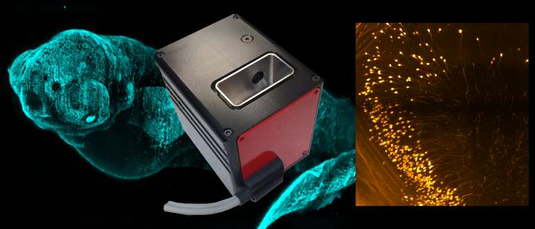
- 3D imaging in light sheet microscopy
- Super-resolution techniques
- Sample preparation
- Intravital imaging and small animal imaging
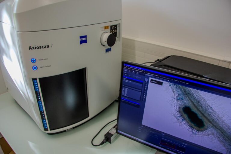
Practical work (60 %) – Carried out in small groups on various equipment at the Toulouse imaging platform (widefield, confocal, multiphoton, SPIM microscopes, etc.)
- Advantages and limitations of the different systems
- Acquisition and analysis of images under the best conditions
Possibility of carrying out the practical work, for educational purposes, on samples brought in by trainees
EQUIPMENT & PERSONNEL
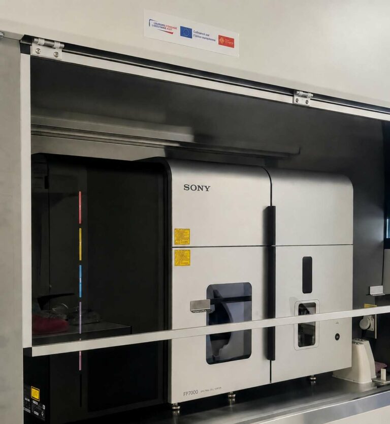
- Wide-field microscopes, confocal microscopes, multiphoton microscopes, CCD cameras, light sheet microscope and macroscope (SPIM), intravital microscope, whole-body fluorescence imaging system
- Researchers and engineers from the following research institutes: Agrobiosciences, Interactions and Biodiversity Research Federation; Toulouse Biomedical Research Federation (SFR BMT)
Contact and registration
- For further information, contact: Jacques Rouquette
- Applications on the CNRS website