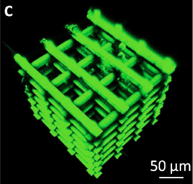


3D architectures are essential tools that, by recreating environments of biomimetic cellular cultures, allow the study of cell growth, proliferation, and differentiation. Researchers from the LAAS (CNRS), in partnership with the TRI-Genotoul platforms of the CPTP and Restore, have managed, using laser engraving performed using multiphoton microscopes, to fabricate self-supporting 3D scaffolds. These scaffolds were then seeded with neural cells and characterized using advanced approaches of 3D fluorescence imaging. The results published in the journal Small The results of Advanced Science news are particularly encouraging.
The technique employed here allows for a much finer manufacturing process (≈ 200 nm) than conventional 3D printing technologies based on fused deposition modeling, inkjet printing, selective laser drying, or the «polyjet» technology.
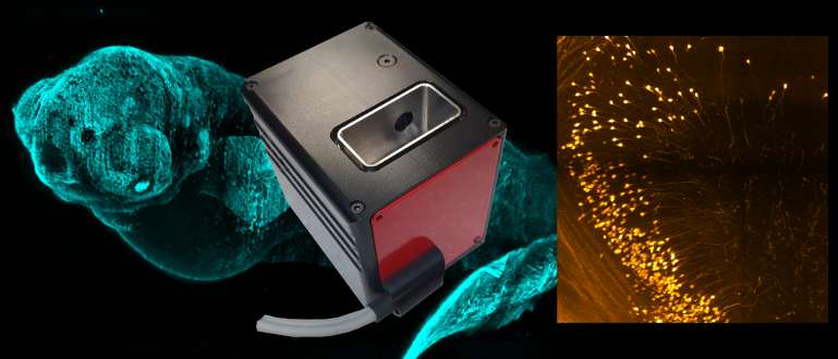
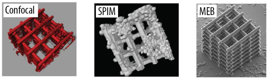
The samples thus created were then studied using scanning electron microscopy (SEM), which revealed the morphology of the cells and the cellular connections around the 3D architecture. The positioning of the nucleus, inaccessible by conventional SEM imaging, is revealed by light sheet fluorescence microscopy and multiphoton confocal imaging. These different techniques have highlighted long neural extensions and optimal cellular colonization around and within the 3D scaffold. These new architectures and characterization techniques open up interesting perspectives for both neural cell culture and observation.