


 |
A very high resolution for observing and analyzing the composition of elementary structures |

|
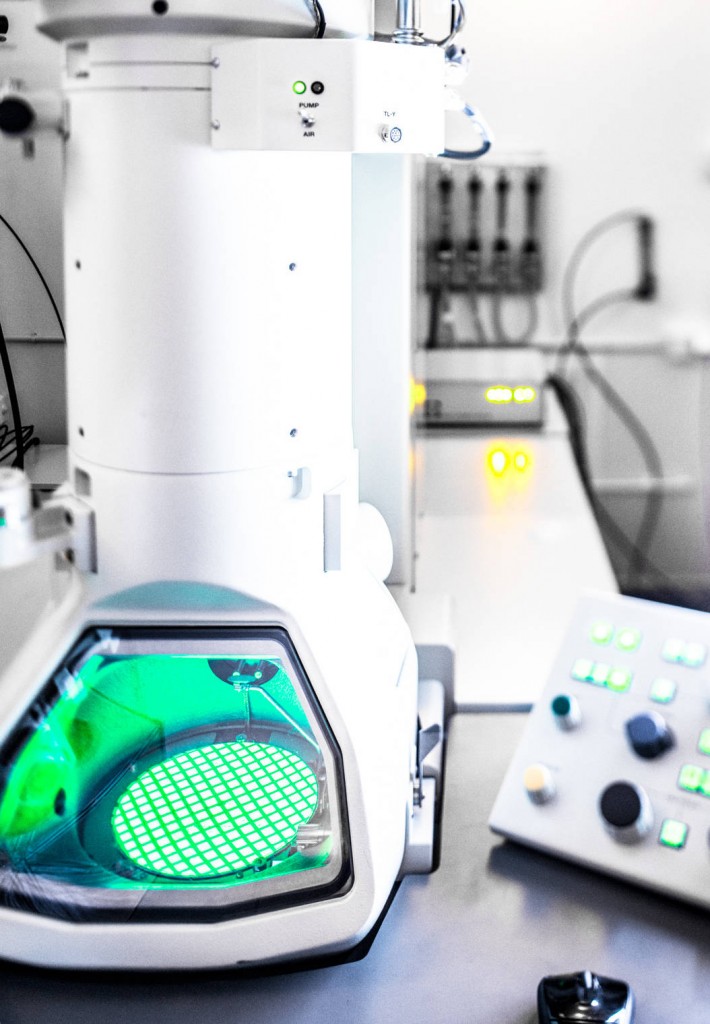
As its name suggests, electron microscopy is not based on photons but on electrons. Transmission electron microscopy (TEM) can therefore achieve a very high resolution (magnification from 50x up to 1.2 million x). A high-energy electron beam is transmitted through a thin sample and the image is formed on a phosphorescent screen coupled to a digital camera. TEM has many applications associated with different preparation techniques, including fine observations of the internal structures of cells, bacteria, viruses, etc.
Services :
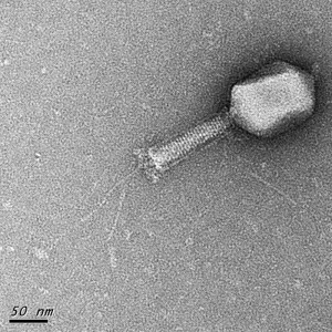
Negative staining of T6 phages – Observation at 100KV, x50000
- Traditional observation in high-contrast and high-resolution mode
- Cryomicroscopy
- 3D observation by electron tomography
- Filtered imaging for analysis of chemical composition
- Sample preparation (chemical fixation, resin embedding, semi-fine to ultra-fine slices, and coloring)
- Cryo-methods
- Manual or automated immuno-labeling
- Image analysis
Access mode:
- With assistance
- In autonomy (after training)
- As Service delivery
TRANSMISSION ELECTRON MICROSCOPES
Name |
Technical characteristics |
Location |
| ThermoFisher Talos Arctica | x 60 to x 1 100 000, equipped for tomography and high-resolution cryomicroscopy. | METi – UPS Campus |
| Jeol JEM 2100 | x 50 to x 1 000 000, equipped for cryomicroscopy, tomography and elemental analysis | METi – UPS Campus |
| Jeol JEM 1400 | x 50 to x 1 200 000 equipped for tomography | METi – UPS Campus |
| MET HT 7700 Hitachi | x 200 to x 600 000, remote control | CMEAB – Rangueil |
Some publications made thanks to these resources:
- Carron C, Balor S, Delavoie F, Plisson-Chastang C, Faubladier M, Gleizes PE, O’Donohue MF; J. Cell Science Octobre 2012, Post-mitotic dynamics of pre-nucleolar bodies is driven by pre-rRNA processing.
- Rath P, Saurel O, Czaplicki G, Tropis M, Daffé M, Ghazi A, Demange P, Milon A.. Cord factor (trehalose 6,6′-dimycolate) forms fully stable and non-permeable lipid bilayers required for a functional outer membrane. Biochim Biophys Acta. Sep. 2013.
- Atomic force and electron microscopic-based study of sarcolemmal surface of living cardiomyocytes unveils unexpected mitochondrial shift in heart failure. Dague E, Genet G, Lachaize V, Guilbeau-Frugier C, Fauconnier J, Mias C, Payré B, Chopinet L, Alsteens D, Kasas S, Severac C, Thireau J, Heymes C, Honton B, Lacampagne A, Pathak A, Sénard JM, Galés C. J Mol Cell Cardiol, 2014, 74, 162-72.
- 5,6-Epoxy-cholesterols contribute to the anticancer pharmacology of tamoxifen in breast cancer cells. Segala G1, de Medina P, Iuliano L, Zerbinati C, Paillasse MR, Noguer E, Dalenc F, Payré B, Jordan VC, Record M, Silvente-Poirot S, Poirot M. Biochem Pharmacol, 2013, 86(1), 175-89.