


 |
Comprehending the volume of objects through TEM |

|
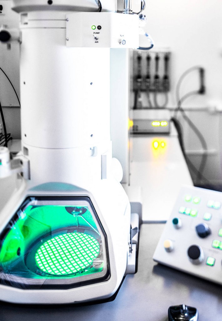
Electron tomography makes it possible to recreate volumes at a nanoscale, by means of a Transmission Electron Microscope (TEM). Just like a medical scanner, images of a given object are made from different angles. The use of imaging software then allows the reconstruction of the sample’s volume and its 3D visualization. The preparation of samples is the same as for observation by TEM.
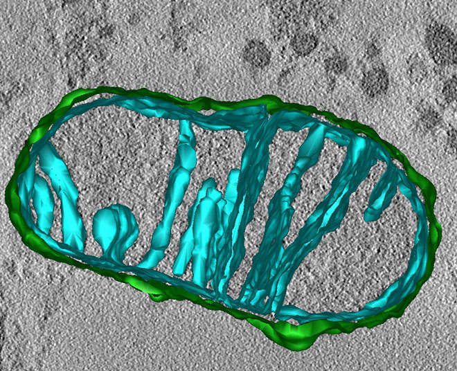
Mitochondria reconstructed by electron tomography
Services:
- 3D reconstruction of objects up to 300 nm thickness
Access mode:
- With assistance
- As service delivery
Transmission electron microscopes (TEM)
Name |
Technical characteristics |
Location |
| ThermoFisher Talos Arctica | x 60 to x 1 100 000, equipped for tomography and high-resolution cryomicroscopy. | METi – UPS Campus |
| Jeol JEM 2100 | x 50 to x 1 000 000, equipped for cryomicroscopy, tomography & electron energy loss filter | METi – Campus UPS |
| Jeol JEM 1400 | x 50 to x 1 200 000, equipped for tomography | METi – Campus UPS |
Some publications made thanks to these resources:
- The path of pre-ribosomes through the nuclear pore complex revealed by electron tomography. Nat Commun., 10(1):497. Delavoie F., Soldan V., Rinaldi D., Dauxois J-Y., Gleizes P-E. (2019)