 |
High-resolution observation and analysis of samples |

|
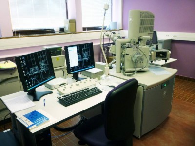
Scanning electron microscopes are used to view the surface of various samples and to observe details on a nanometer scale. Moreover, control of the pressure in the microscope chamber and the cryo-preparation module offers the ability to observe dry, hydrated or frozen samples. Among the many possibilities offered by this resource, cryo-fracture can be used to observe the interior of samples and X analysis can determine and quantify the atomic species present on its surface.

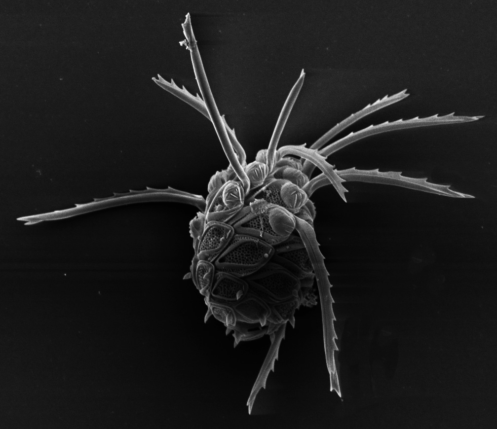
Phytoplancton – CMEAB
Services :
- Traditional observation in high vacuum mode
- Observation of hydrated samples in environmental mode
- Observation of non-conductive samples in variable pressure mode
- Cryomicroscopy (cryofixation in slush nitrogen, sublimation, cryofracture)
- Sample preparation: CO2 critical point, metallization (Pt, AuPd, Cr)
- Immuno-labeling (immunogold)
- X analysis for analyzing the chemical composition
Access mode:
- With assistance
- As service delivery
SCANNING ELECTRON MICROSCOPY
Name |
Location |
| S.E.M. ESEM Quanta 250 FEG | CMEAB – Rangueil medecine faculty |
Some publications arising from the use of these tools:
- Deciphering the elusive nature of sharp bone trauma using epifluorescence macroscopy: a comparison study multiplexing classical imaging approaches. Capuani C, Rouquette J, Payré B, Moscovici J, Delisle MB, Norbert Telmon, Guilbeau-Frugier C. International Journal of Legal Medicine, 2013, 127, 169-176.
- Scanning electron microscopy of Haemonchus contortus exposed to tannin-rich plants under in vivo and in vitro conditions. Martínez-Ortíz-de-Montellano C1, Arroyo-López C, Fourquaux I, Torres-Acosta JF, Sandoval-Castro CA, Hoste H. Exp Parasitol, 2013, 133(3), 281-6.


