


 |
Counting photons to quantify spatial proximity between molecules |

|
These multi-photon confocal microscopes are equipped with FLIM imaging modules to produce lifetime images. In biology, this type of module is mainly used to measure the spatial proximity of membrane receptors previously labeled with fluorochromes. More generally, FLIM imaging is a prerequisite for any comparison of fluorescence intensities in a confocal image and therefore the analysis of spatial proximity of molecules.
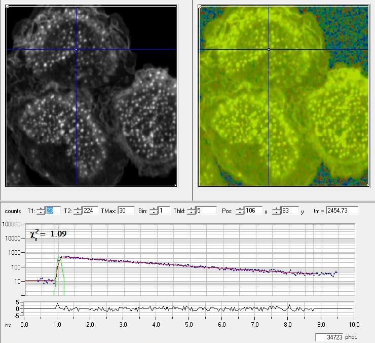
Left: conventional fluorescent image in black and white to show the position in the sample – Right: lifetime image – Graph: lifetime by photon counting
Services:
- XYZT acquisition, multi-color, spectral separation
- Interactions between molecules (FLIM function)
- Multi-labeled images: 2D to 5D + DIC
- Evaluation of molecular traffic between compartments (FRAP)
- Fluorescence anisotropy measurements
Access mode:
- With assistance
- In autonomy (after training)
- As collaborative project
- As service delivery
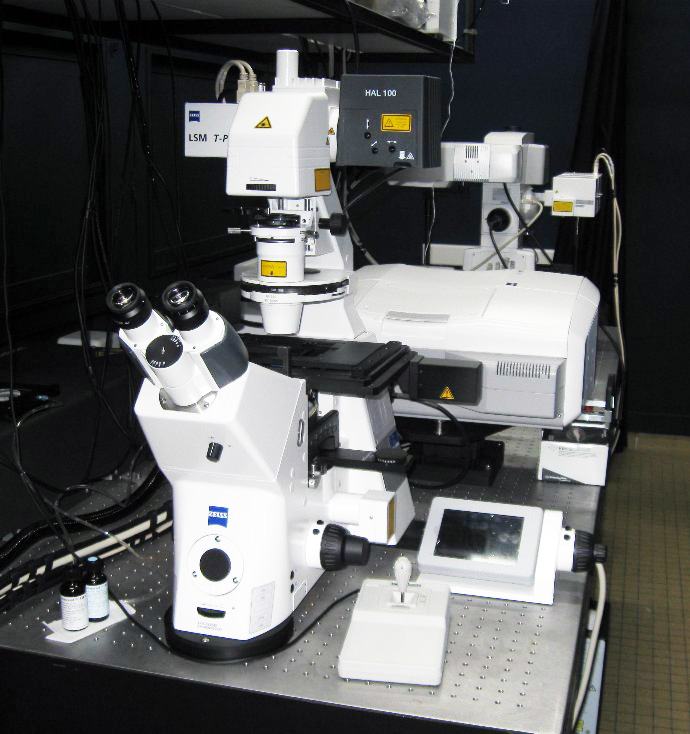
Available Confocal FLIMs
Nom |
Caractéristiques techniques |
Lieu |
| Multi-photon confocal microscope FLIM 7MP | Mounted on an upright microscope, thermoregulated | IPBS – Rangueil |
| Multi-photon confocal microscope FLIM 7MPFLIM LSM710 | Mounted on an inverted microscope, thermoregulated and CO2 controlled chamber | IPBS – Rangueil |
| Mono-photon confocal microscope LEICA SP8 SMD | Mounted on inverted microscope | FR3450 – Campus INRA Auzeville |