 |
Observing living specimen in 3D and in depth |

|
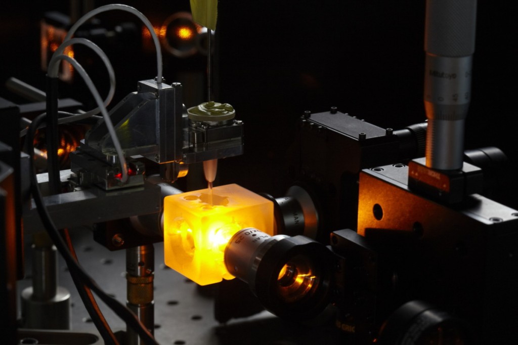
The work of R&D performed at Restore led to develop microscopes based on light sheet excitation or SPIM (Selective Plane Illumination Microscope). This technique not only enables fast optical sectioning of living specimen in 3D and in depth but also offers the possibility of multiview acquisitions, opening new perspectives in biological observation. On the same principle, the MacroSPIM, which is also based on light sheet excitation and built thanks to IRB partnership, makes it possible to view tissues and whole organs (after they have been made transparent) by rapid optical sectioning, again in 3D and in depth. The fields of application are numerous and can be used on both animals and plants.
Services :
|
Observation, in depth, of mitosis by SPIM microscopy (in red: cells in mitosis; in blue: cells in interphase)
|
Modes of access:
|
Booking
Name: |
Lieu : |
|
| LIGHT SHEET MICROSCOPES (SPIM) | Equipped with three continuous lasers (491, 532, 595 nm) Thermoregulated and CO2 controlled chamber. Small samples (e.g. cells, tissues …) |
Restore – Paul Sabatier University UT3 |
| MACRO-SPIM | Equipped with four continuous lasers (405, 488, 561, 642 nm). Samples of several cm3 in size (e.g. organs …) | Restore – Paul Sabatier University UT3 |
| Light Sheet Z7 (Zeiss) | Equipped with four continuous lasers (405, 488, 561, 638nm). Samples from several μm to cm. Thermoregulated controlled chamber. | Restore – Paul Sabatier University UT3 |
Some movies made thanks to these tools
With SPIM |
|
A journey into a spheroid – Restore |
Cell in mitosis – Restore |
With MacroSPIM |
|
|
Mouse gut – Restore |
|
|
Mouse gut – Restore |
Plant – Restore |
More informations
To learn more on last SPIM devloppements made by Restore teams, please visit R&D imaging webpage.


