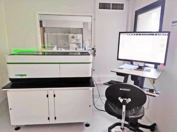
TRI-Genotoul brings its expertise in the fields of medical research, basic sciences, cancer and rejuvenation, and agro-biology. In addition, eight R&D teams (LAAS, CBI, IPBS, IRSD and IMT) complete the node and support the facility to drive continuous improvement in expertise and technologies.
LAAS – Engineering for Life Sciences and Applications (ELiA)
Head: Laurent Malaquin
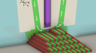
- Expertises:
- 3D printing and 3D Bioprinting applied to biodetection
- Development of 3D microenvironment as models for cell culture and cancer study.
- Topics: Microenvironement for cell culture, tissue engineering, cancer diagnosis, microphysiological systems.
- Skills: Microfluidics, 3D printing, Bioprinting, Self assembly, Biopatterning
 LAAS – Micro-Nanofluidics for Life and Environmental Sciences
Head: Pierre Joseph
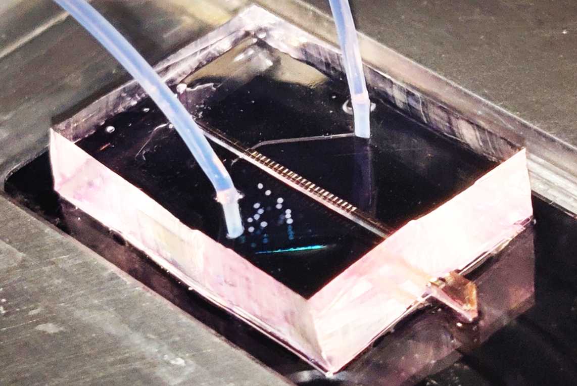
Development of microfluidic confining culture chambers, in order to study the impact of spatial confinement. Adapted to mechano-biology and allowing well-defined microenvironment.
 MCD-CBI: Chromatin and gene expression
Head: Kerstin Bystricky
The team develops original systems to fluorescently label and track DNA (ANCHOR technology – NeoVirtech SAS) in real time at nanoscale resolution to understand physical principles underlying regulation of gene expression and DNA repair, in cellular plasticity and tumorigenesis.
 IPBS – Phagocyte architecture and dynamics
Head: Christel Vérollet & Renaud Poincloux
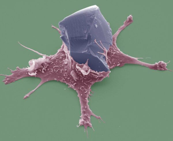
This R&D team combines cutting-edge techniques in optical and electron imaging, material science, cell mechanics and intra-vital imaging to elucidate how phagocytes, in particular macrophages and osteoclasts, interact with the extracellular matrix, to decipher the mechanisms of macrophage 3D migration and phagocytosis, and investigate how HIV-1 manipulates phagocyte cell-to-cell spread.
MCD-CBI: Dynamics and disorders of ribosome synthesis
Head: Pierre-Emmanuel Gleizes
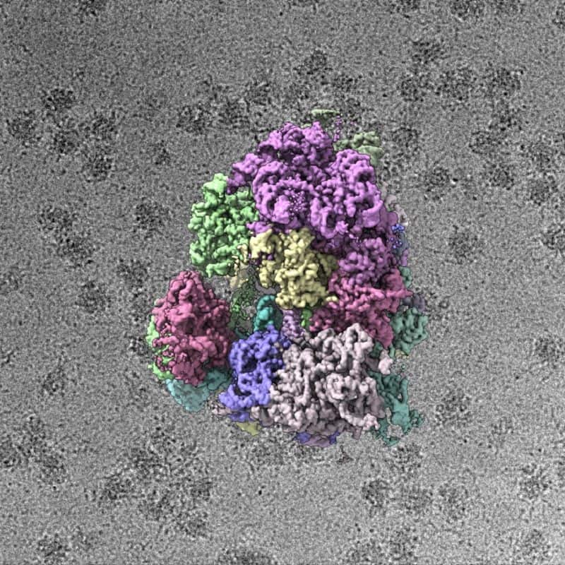
3D-electron microscopy approaches and correlative microscopies at different scales, from single molecules to tissues. Strong expertise in molecular imaging by cryo-EM and single particle analysis
MCD-CBI : OncoRib – Ribosomes in Normal and Pathological conditions
Head: Celia Plisson-Chastang

Our team develops cryo-electron microscopy (cryo-EM) coupled to image analysis approaches such as single particle analysis and cryo-electron tomography. We aim to characterize the 3D structures and spatial organization of molecular machines like ribosomes and chaperones, to understand how they function in normal and pathological conditions.
IRSD: Gut Protease Signals
Head: Nathalie Vergnolle
Expertise in 3D imaging of live and fixed organoids. Drug screening tests using morphological characterization of organoid cultures as readouts. Organization of an open platform for Organoid development.
 CBI / TRI: R&D LITC platform
Head: Thomas Mangeat
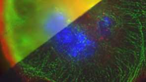
Dévelopment of the RIM technology (Random Illumination Microscopy). Development of low cost integrated optical technology.
 MCD-CBI: MAthématiques pour l’iMagerie BiOlogique (MAMBO)
Head: Pierre Weiss
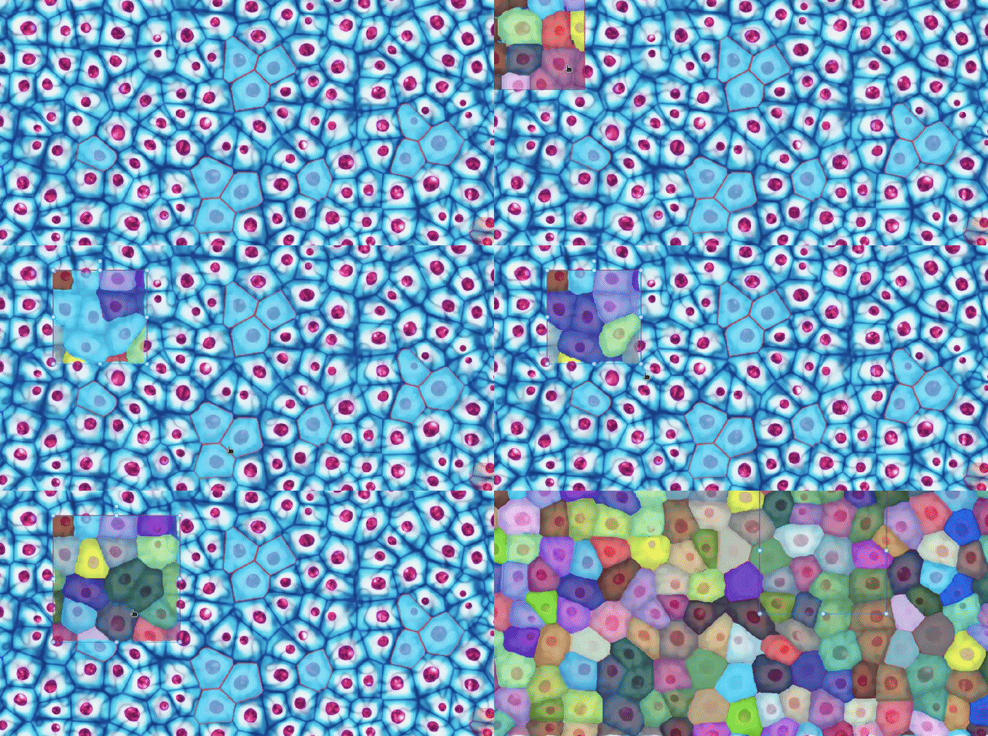
The MAMBO team (MAthématiques pour l’iMagerie BiOlogique) is specialized in mathematics (optimization, learning, approximation, inverse problems) and in imaging (reconstruction and analysis). It has 2 main objectives:
- Participate in the improvement of microscopy techniques (e.g. RIM, TIRF, SMLM, …) by refining their mathematical modeling and by building efficient reconstruction algorithms.
- Participate in the analysis of the resulting images by developing (or using) tools to improve the quality of segmentation or classification in large volumes of data. In particular, it develops new artificial intelligence models to achieve its goals.


