 |
For high throughput observation and analysis of cell populations |

|
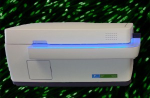
High throughput imaging combines instruments dedicated to the acquisition, measurement and analysis of the cell contents from various microscope slides or plates (up to 384 wells). They come together in a wide-field automated fluorescence microscope used to analyze fixed or living cells under a controlled environment. These intuitive resources offer the possibility to analyze multiple structural and functional cellular parameters in parallel. A dedicated server enables the storage, management and analysis of data remotely.
Services:

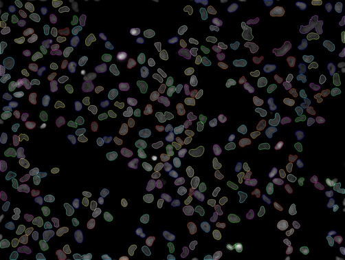
These instruments automatically detect cells in order to proceed with analyses of the cell contents
- Confocal or wide-field XYZT acquisition, multi-color fluorescence or transmitted light
- Live or fixed samples
- Up to 384 samples in one pass
- Analysis of fluorescence parameters
- Analytical capabilities for many research and screening (HCS) applications: cell biology, immuno-labeling, cytotoxicity, pharmacokinetics, cancerology, etc.
- Management, data storage and “deported” analyses
Access mode:
- With assistance
- In autonomy (after training)
- As service delivery
MULTIPARAMETRIC HIGH THROUGHPUT IMAGING AND ANALYSIS
Name |
Characteristic techniques |
Location |
| Operetta – High Content Imaging System PERKIN ELMER |
20x & 40x confocal spinning disk mode transmission mode Pixel size 6,45×6,45µm Thermoregulation (37°C & CO2 from 3 to 8,5%) |
LBCMCP – UPS Campus |
| THERMO CELLOMICS ARRAYSCAN® VTI HCS Reader | From 5x to 40x Transmitted light module Pixel size: 6.45×6.45µm Thermoregulation (37°C & CO2 from 0 to 10%) |
Restore – Oncopole |


