


 |
For generating homogeneous populations and analyses without contaminants |

|
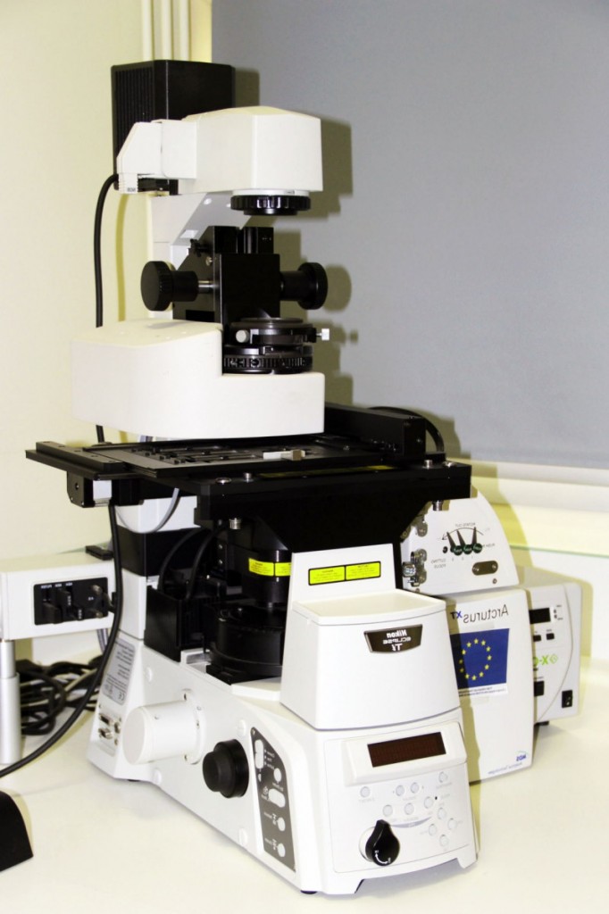
The laser microdissector ion system is not an imaging technique per se, and it must be coupled to a microscope. It can then be used to observe tissue slices, cut the areas to be sampled (cell layer, fungus, etc.) and collect them on a capsule in order to conduct specific biochemical analyzes. Tests may thus be conducted on a few cells of interest, without alteration of the information by the remaining tissues from which they were extracted. This technique makes it possible to perform qPCR, dosage, proteomics, sequencing and even microRNA analyses.
Services:
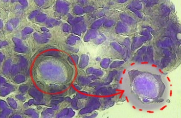
Targeted removal of a particular cell within a tissue
- Samples microdissection (10 to 200 µm thickness) in fluorescence and in bright-field microscopy
- Capture of microdissected samples with a thermosensitive CAPS membrane activated by an infrared laser; possibility to also use a UV laser to cut the area to be sampled by first laying down the sample on a PEN membrane-covered microscope slide to get rid of the object’s adhesion on the microscope slide
- Special CAPS (HS) that limit their contact with the sample, enabling the sampling of single cells
- Coupled with an inverted microscope allowing image acquisition of the slide with mosaicking for quick identification of some area of interest
Micro-dissectors
Name |
Technical characteristics |
Location |
| Microdissector Arcturus XT | Coupled to Nikon Eclipse inverted microscope | FRAIB – Campus INRA Auzeville |